Common Foot Injuries of the Equine Athlete
Updated:
December 1, 2023By Dr. Billy Hodge
We ask a lot of our four-legged friends as today’s horses compete throughout most of the year. Whatever the horse’s primary job — from dressage to trail riding and reining to show jumping, the feet are the most common source of lameness. With the advent of preventative drugs such as Legend®, Adequan®, and Pentosan EQ™ we can improve the longevity of our horses’ athletic use, but unfortunately injuries still occur.
When confronted with lameness, your veterinarian will take a thorough history and perform a careful examination utilizing their eyes and hands, their experience, and most importantly, a lameness exam, to make a diagnosis. Most cases will be quickly diagnosed, while others will require more intensive tests requiring diagnostic blocking (use of local anesthetic or numbing agents to localize the lameness to a specific region). The veterinarian may suggest further imaging with radiography (x-rays), ultrasound, bone scan, and/or magnetic resonance imaging (MRI) to diagnose the problem.
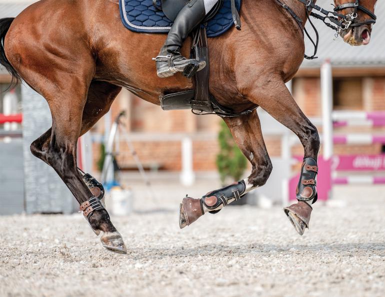
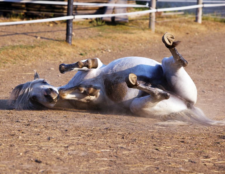
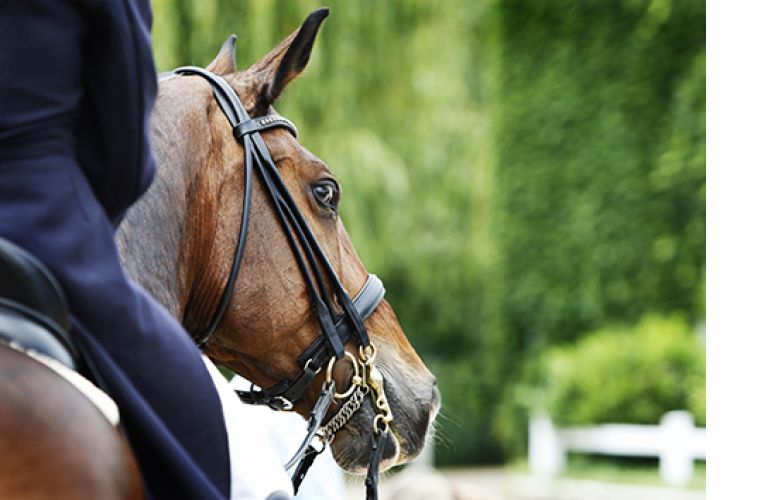
Acute pain during or immediately following work can mean a diagnosis of soft tissue injury. Photo: Dreamstime/CustomPosterDesigns
A definitive diagnosis allows the veterinarian to treat your horse effectively with the best chance at a complete return to pre-injury level. Here are the most common sources of foot lameness we see at our clinic (which may differ from other clinics depending on equine disciplines served):
- Subsolar abscess
- White line Disease
- Laminitis
- Joint inflammation (coffin joint synovitis/arthritis)
- Foot bruise (including bone bruise)
- Soft tissue injury (examples): Deep digital flexor tendonitis; Collateral ligament injury
- Navicular disease and navicular bursitis
- Fracture
Hoof abscesses are common after periods of wet weather followed by dry weather causing small, micro-cracks in the hoof wall. These cracks subsequently allow bacteria to enter the hoof, only to create an abscess. There is usually moderate to severe lameness, significant increase in digital pulses, heat (compared with the opposite foot), and sensitivity to hoof testers. A hot poultice worn for multiple days followed by careful paring of the sole by your vet or farrier will allow the abscess to drain. Occasionally the abscess may burst from the coronary band, in which case antibiotics may be required.
A hoof abscess (small hole at toe) results when small cracks in the hoof wall allow secondary bacteria or a fungal infection to enter the hoof. Photo: Shutterstock/Chelle129
White line disease (WLD) is caused by abnormal stresses on the hoof wall that cause the hoof wall to separate from the underlying laminae. This leads to a hollow, weak, and destabilized hoof wall. A secondary bacterial or fungal infection can then take hold in the abnormal hoof wall. We usually diagnose WLD based on a careful examination of the hoof wall, then aggressively but carefully remove the damaged wall and utilize hoof acrylics (with antibiotics) and corrective shoeing to reshape the foot.
Related: Should My Horse Be Barefoot or Shod? It Depends
Laminitis is a frustrating and crippling disease that can affect any horse. Laminitis is inflammation and eventual failure of the sensitive laminae of the feet that attach the hoof wall to the pedal bone. This results in rotation or sinking of the pedal bone within the hoof capsule. Laminitis can be caused by several different mechanisms including metabolic, mechanical, or it can be secondary to endotoxemia. Metabolic causes are seen in horses in early spring when the grass has high levels of easily fermented starches, or in some horses that are predisposed to metabolic disease (equine metabolic syndrome). Mechanical laminitis is caused by excessive weight-bearing on one leg when the opposite leg is injured. The third trigger is severe illness that causes endotoxemia such as colic, colitis, and pneumonia.

The laminitis hoof wall (left) compared to the healthy hoof wall (right). Photo: Clix Photography
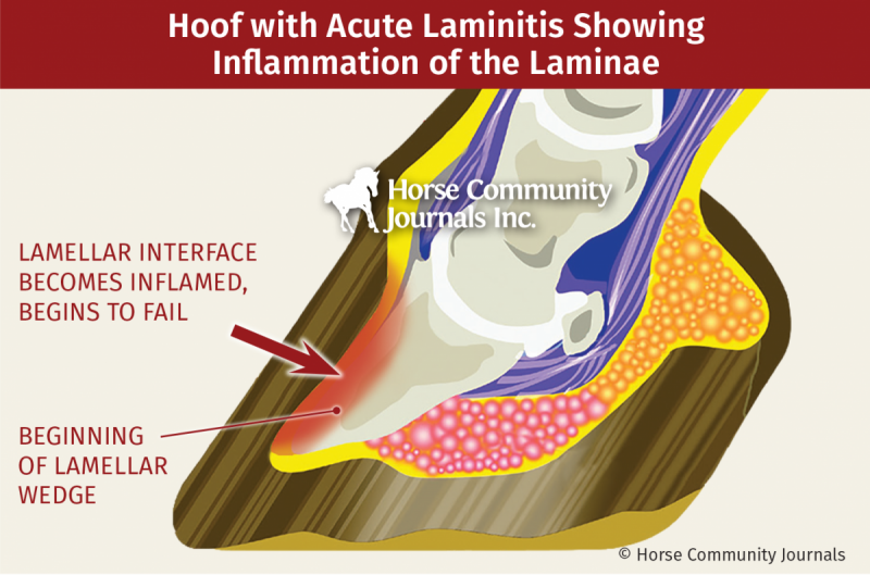
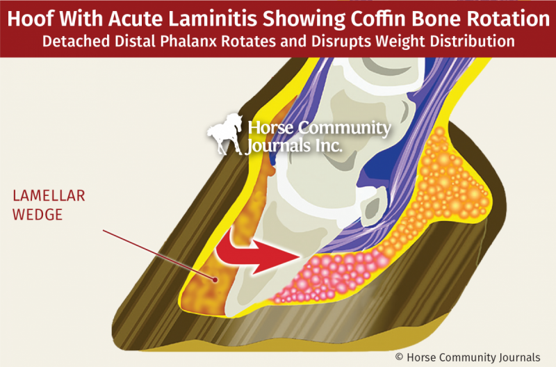
Chronic laminitis can be prevented by early recognition, diagnosis (x-rays), and treatment. Treatment is to remove the inciting cause, ice the feet aggressively in the acute stage, provide pain relief, and support the foot using deep bedding and special shoes. The goal is to halt the progression of laminitis. Together with a good farrier and careful control of pain we have been successful in returning horses to their athletic careers.
Related: Finding the Hoof Care Balance - Top Tips & Myth Busters
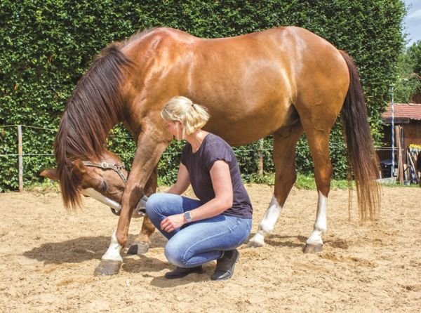
Foot bruises are commonly seen the day after a hard training session. Soft tissue bruising resembles a hematoma with bleeding into the soft tissues of the foot between the pedal bone and the hoof wall. The most common sign is mild to moderate lameness with reaction to hoof testers. There is usually a pulse in the foot and horses can be sound in one direction on the lunge and lame in another direction. There is significant improvement with rest and anti-inflammatory medication, but recurrence after removal of medication is common. A nerve block will pinpoint the foot as the source of lameness, although radiographs may be normal.

Hoof testers are a valuable diagnostic tool to gauge sensitivity and pain response. Photo: Pam MacKenzie
There can also be actual bruising of the pedal bone, which is a more serious injury. This can be diagnosed on bone scan where the bone has an increased uptake of signal or on MRI where bone edema of the pedal bone (P3) or pastern bone (P2) can be seen. Horses usually require a period of rest of one to three months, and may require medication such as aspirin, Osphos (clodronate Disodium), and low dose bute (phenylbutazone).
Joint inflammation or degenerative joint disease occurs due to the high levels of stress loads during exercise and by general wear and tear. Joints can become painful for many reasons, but usually this occurs secondary to trauma, excessive twisting, and/or landing and turning suddenly. This causes inflammation and results in heat, joint swelling, pain on flexion, and lameness. The most common diagnostic test is to administer five ml of a local anesthetic into the joint and quickly reassess the lameness on the lunge; there should be relief within five to ten minutes. X-rays are usually negative, although some cases may have mild changes at the edge of the joint called osteophytes, suggestive of degenerative joint disease. Treatment is easy and effective for months to years when horses are allowed a period of five to seven days of rest after injections with hyaluronic acid (HA) with or without steroids. We also now commonly use IRAP (interleukin-receptor antagonist protein) or platelet rich plasma (PRP) in order to provide anti-inflammatory and/or regenerative therapy in conjunction with remedial shoeing to improve long-term success.
Related: Osteoarthritis - Keeping Our Aging Horses Sound

A veterinarian performing diagnostic blocking to pinpoint the source of lameness. Photo: Clix Photography
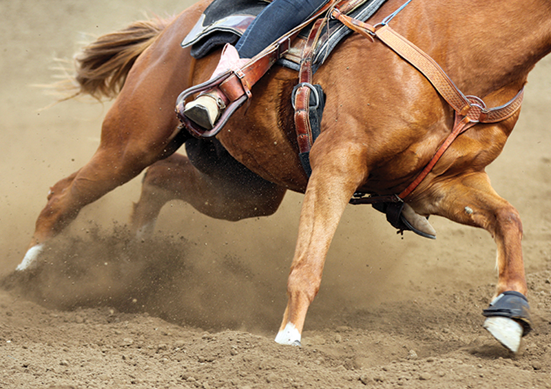
Soft tissue injuries usually present as acute lameness during work or immediately following competition. The deep digital flexor tendon in the foot acts to help flex the foot and attaches to the bottom of the pedal bone within the hoof capsule. The deep digital flexor tendon can be strained or torn, and when this occurs we see acute lameness. The tendon cannot be palpated in the foot and tendon injuries in the foot cause lameness without heat, but deep palpation in the heel bulbs or just above may cause horses to react. Horses usually show significant lameness in a straight line. Horses will be negative to hoof testers, but there will be pain following flexion tests with increased lameness. Both nerve and joint blocks will be very important in fully localizing the lameness. Ultrasound and MRI are essential in the diagnosis of these cases and treatment depends on the structure injured and the severity of injury. Treatment can include rest, anti-inflammatory medication such as nonsteroidal anti-inflammatory drugs (NSAIDs) or corticosteroids, remedial shoeing, IRAP, PRP, stem cells, or surgery coupled with a specific rehabilitation program.
Related: Treating Navicular Disease with Farriery
Collateral ligaments of the coffin joint are another soft tissue commonly injured in the foot. These ligaments attach the pedal bone to the short pastern bone and stabilize the foot during exercises that involve changing direction quickly. Mild strain or tear in these ligaments is somewhat similar to rolling your ankle, as the coffin joint can become unstable after the injury. Treatment varies depending on the severity and degree of bone involvement, but can include IRAP, extra corporeal shock wave therapy (ESWT), rest, treatment of concurrent coffin joint inflammation, and even PRP injection into the lesion.
Navicular disease and navicular bursitis are now recognized in all disciplines and all breeds. The navicular bone is a small bone in the back of the foot that sits between the pastern and pedal bone, allowing a smooth gliding surface for the deep digital flexor tendon to pass behind. A small fluid-filled sac called the navicular bursa sits between the navicular bone and deep flexor tendon. Because of the close proximity of these structures, they are usually all considered when looking at lameness localized to the back of the foot.
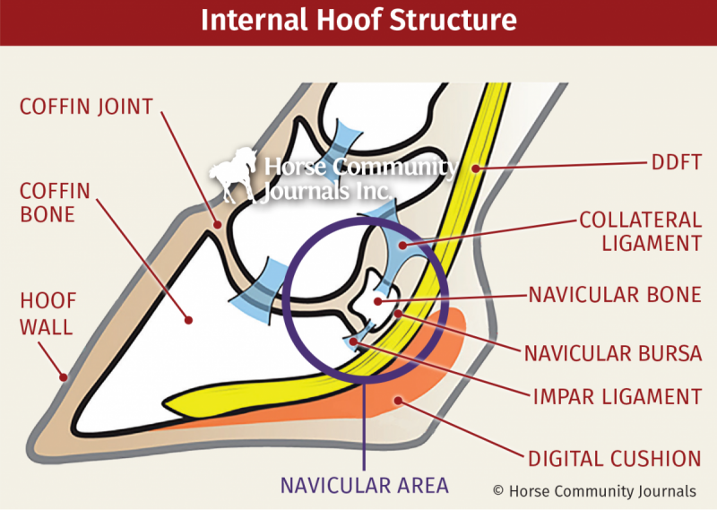
Most horses will present with a low-grade lameness, which waxes and wanes during the competition season. The lameness workup will localize the lameness to the back of the foot/heel region with no clear soft tissue injury seen after ultrasound. In some cases obvious changes can be seen on radiographs, but this may not be the case for younger horses that have been in work only a short time. MRI can also pick up bone edema (swelling) that cannot be seen on x-rays. Navicular disease cannot be cured but it can be managed with remedial farriery (heart-bar or egg-bar shoes), NSAIDS (bute, banamine), Isoxsuprine (a vasodilator), periodic rest, Tildren/Osphos (bone pain modification), ESWT, acupuncture, and navicular bursa injections.
Fractures are less common, and a quick and accurate diagnosis often allows a full return to function. Horses will often present with an acute lameness and radiographs will show a fracture line. Many fractures heal in three months with rest, but some may require surgery. We commonly use remedial shoes and casts in combination with rest or surgery.
Related: Serviceably Sound - What Does It Mean?
Related: 10 Things You Might Not Know About Horse Hooves
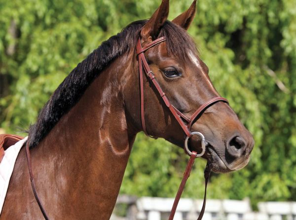
Main Photo: Shutterstock/El-Ka
Advertisement
Related Articles


Advertisement
Featured Stories


Advertisement