
Evaluating Equine Lameness Using New Technology
Updated:
January 8, 2024By Tania Millen, BSc, MJ
Keeping horses sound is one of the most difficult and important aspects of horse sport. Given that horses have an uncanny ability to injure themselves, at some point every rider or horse owner will need their veterinarian to conduct a lameness evaluation.
Assessing the nature and severity of lameness includes discussing the horse’s history, palpating the horse’s musculoskeletal system, checking hooves, and observing the horse at walk and trot before flexing joints and deciding whether to freeze or block nerves in the legs, take x-rays, perform ultrasound or complete magnetic resonance imaging (MRI). Now, new technology — Equinosis Q with Lameness Locator® — can help veterinarians confirm their hunches. It’s a game changer for correctly diagnosing injuries and developing treatment plans, potentially saving horse owners hundreds of dollars. However, the horse isn’t just hooked up to a machine which spits out a diagnosis of “the horse is lame on the left front due to an arthritic knee,” for example. Far from it. Interpretation is needed and for that reason, the system is sold only to licenced registered veterinarians and not every equine veterinarian has one.
Dr. Connie Fancy, who owns and operates Fancy Veterinary Services in Claresholm, Alberta has been conducting lameness evaluations in performance horses for 28 years. She invested in the technology and describes how she uses it as part of lameness evaluations. She also explains that every lameness evaluation starts with a general overview.
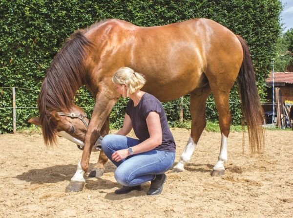
For prepurchase exams, assessing lameness, and monitoring therapy and rehabilitation, the Lameness Locator® provides reliable data that assists with veterinary diagnosis and informs training and management decisions. Photo: Dreamstime/Kseniya Abramova
Related: Equine Lameness Evaluation
1. Hands on the horse
“Looking at and feeling the horse are the most important parts of any lameness assessment,” says Dr. Fancy. “I always start with that because you can feel and see irregularities and asymmetries. The horse will tell you if there are areas that are painful. Even if it’s a joint or bone that’s sore, the muscle over top will be tense.”
After observing and palpating the horse, it’s time to move on.
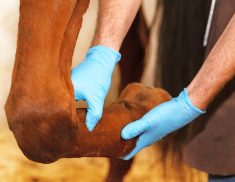
2. Assess the hooves
“Ninety percent of lameness either starts in the feet or becomes a problem in the feet,” says Dr. Fancy. “So, I look at the angles of the pastern, the balance side-to-side, and how the hoof is trimmed or shod. I often see differences in hoof balance or abnormal growth due to lameness and that suggests an issue in that leg or maybe a secondary issue.”
Every hoof is also checked with hoof testers to determine whether there’s pain in the feet.
Related: Evaluating Lameness: An Inside Look
Related: Unravelling the Mysteries of the Pre-Purchase Exam
3. Observe the walk
“Walking in a relaxed fashion on a serpentine pattern tells me a lot about whether the horse is bearing weight symmetrically or whether it’s dragging one foot or more than one foot,” says Dr. Fancy. The horse must walk quietly so that its pain isn’t masked by adrenalin.
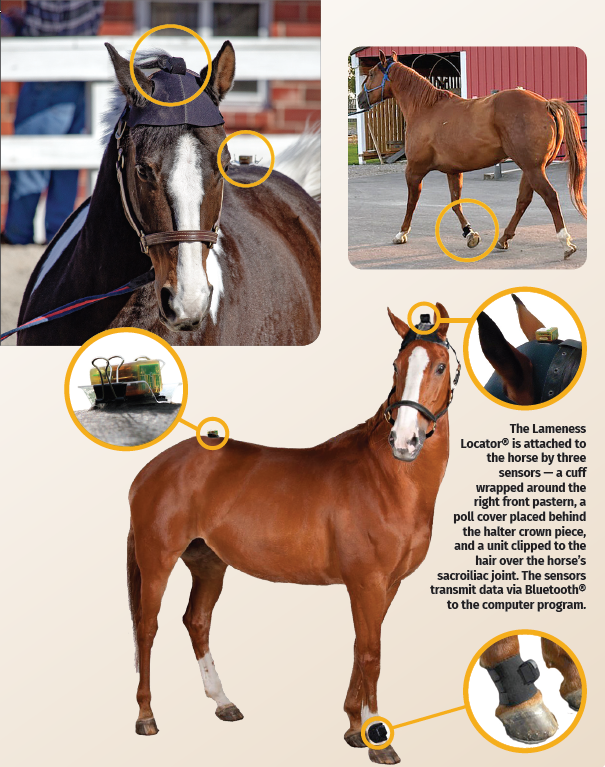
4. Attach Equinosis Q with Lameness Locator®
After these three basic assessments, the Lameness Locator® is attached to the horse. A cuff is wrapped around the right front pastern, a poll covering is placed beneath the halter crown piece, and a small unit is clipped to the horse’s hair over the sacroiliac joint. These three sensors relay information to a computer program via Bluetooth® so no wires are needed. However, a stationary horse doesn’t exhibit many signs of lameness, so the horse is trotted in straight lines in-hand on asphalt to allow the technology to ascertain readings. After that, Dr. Fancy continues with her lameness evaluation while the Lameness Locator® collects data.
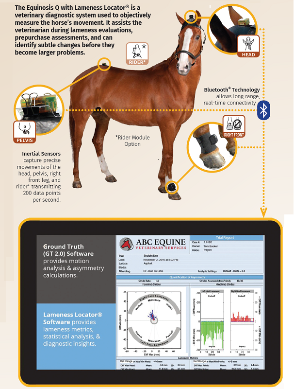
Images courtesy of Equinosis.com
“I pay very little attention to it until the very end of the assessment,” says Dr. Fancy. “I just continue watching the horse.”
How does it work? When a horse feels pain, it places less weight on the correlative leg. The system collects data and generates reports from head and pelvic height measurements, which indicate symmetry differences between how the horse bears weight on each leg. The system also compares how the leg joints are used when each hoof lands (impact), moves though a stride (mid-stance), and leaves the ground (push off). By comparing this data with typical patterns horses display when they have pain in front or hind legs, the system can pinpoint primary, secondary, and compensatory lamenesses, plus distinguish between soft tissue and joint or regional lamenesses.
Although it sounds like magic, for decades horses have been wired up to computers while galloping on treadmills or trotting down long lines of pressure-measuring plates. What’s new is the wireless package coupled with highly evolved software, which uses up-to-date knowledge about equine biomechanics to provide precise data quickly.
Images courtesy of Equinosis.com
Prior to purchasing the technology, Dr. Fancy had to complete a two-week online training course with tested modules.
“I was surprised how extensive the training was,” says Dr. Fancy. “It started with the basics of lameness evaluation, and then got into the physics of the data, data analysis, how the machine works, and how to distinguish different lamenesses.
“I thought it would only be useful for tricky two-legged lameness or mild lameness where it’s difficult to see whether the horse is more sound after being nerve blocked,” says Dr. Fancy. “But I’ve had it for a year and use it on all lameness evaluations and prepurchase assessments now. It’s invaluable.”
Related: Serviceably Sound Horse - What Does It Mean?
Related: Fear Testing of Foals May Help Increase Safety
During an initial lameness exam, the technology provides concrete numbers that can then be utilized during future assessments. For example, if a rehabilitated horse requires re-assessment after six months to determine whether it’s sound, the initial readings allow comparison, reducing the need for the veterinarian to remember what they saw.
“I used to try to explain the lameness to myself in my medical notes, but this is so much more precise,” says Dr. Fancy.
She also says that the Lameness Locator® can identify subtle changes before they become problems, allowing riders to make changes to their programs or initiate treatments, thereby reducing competition downtime or the development of more severe injuries.
5. Observe trot
While the Lameness Locator® collects data, Dr. Fancy watches the horse trot slowly in hand on the asphalt to form her own opinion. Next, the horse is lunged both ways on suitable footing.
“If there’s a mild lameness in either a front or back leg, it will show up more regularly on the lunge because there’s increased weight-bearing on either side,” says Dr. Fancy. “Once we’re finished lunging I have a fairly good overall impression of what the horse is doing. At that point, unless it’s a very obvious lameness, we usually start flexion testing.”
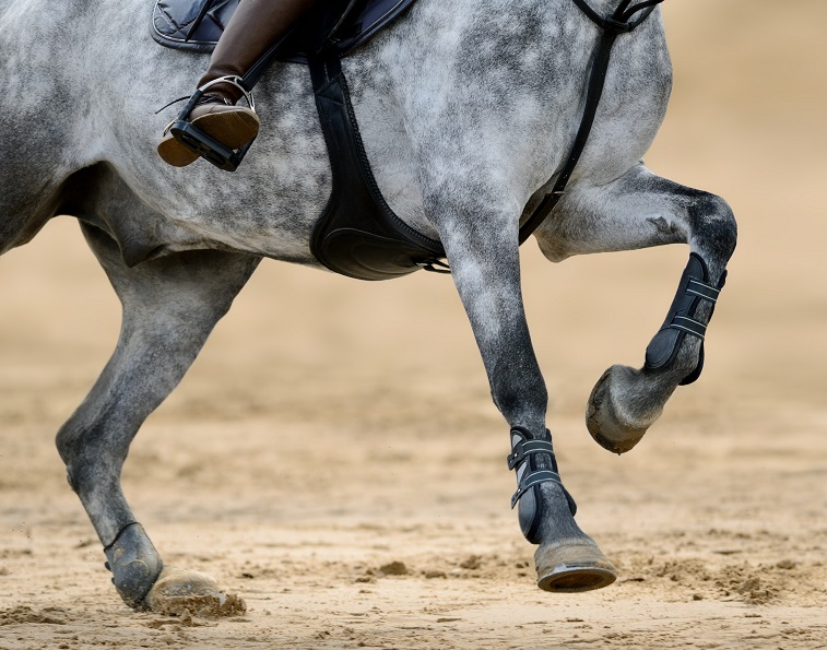
6. Conduct flexion tests
Every veterinarian has a typical pattern they follow for flexion tests, usually starting with the left front leg, and flexing the lower joints (fetlock, pastern, hoof) first. The higher joints (knee, elbow, shoulder) generally follow. However, the pattern depends on the suspected lameness.
“I don’t flex the leg that I think is causing the primary lameness first, because then everything else is meaningless,” says Dr. Fancy. “I will do that one last, so I can identify any secondary or compensatory lamenesses from the other legs. Generally, if you have a front leg lameness, your diagonal hind leg will be involved. If you have a hind leg lameness, the same side front leg will be involved.
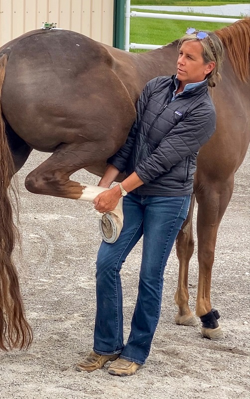
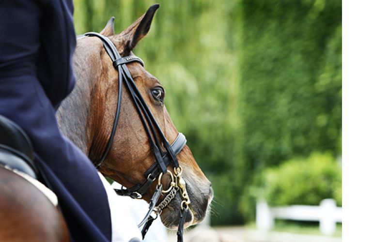
Flexion testing to assist in identifying the source of pain during a lameness evaluation. Photo courtesy of Equinosis.com
“You have to be careful with flexions,” says Dr. Fancy. “If there’s a tendon strain or tear, you have to be careful not to do more harm. Also, if you’re not sure where the lameness is and you keep going back and redoing flexions, then the horse may become sorer than it was before.
“That’s one of the big reasons I find the Lameness Locator® helpful,” says Dr. Fancy. “It speeds up the flexion process and reduces the time it takes to figure out lameness issues.
“Often, I don’t split up the flexions anymore,” she says. “I do a full leg flexion rather than the lowest, middle, and upper joints separately. Once I have a general picture of the lameness and look at the readings, then I have an impression of whether it’s due to soft tissue injury or a joint. At that point, I’ll often go to lameness blocks or freezing sections of the legs. And then localizing the area of pain gets very cut and dry.”
Related: How to Shop for a Horse
Related: Recognizing and Managing the Club Foot in Horses
7. Nerve blocks
Freezing the legs involves injecting local anaesthetic into specific nerves so that the horse can’t feel the area below that point. The anaesthetic “blocks” the pain, so that the horse then trots sound, providing the veterinarian a precise location for the source of the pain. Once nerve blocks have localized the lameness site, the veterinarian will decide whether to x-ray or ultrasound, depending on whether the source of the pain is likely soft tissue, joint, or bone.
8. X-rays, ultrasound, magnetic resonance imaging
“I use ultrasound every day, multiple times a day for lamenesses, and not always for soft tissue concerns,” says Dr. Fancy. “Sometimes I don’t even get the x-ray machine out if I think it’s a joint. The ultrasound machine provides more information on the smoothness or roughness of boney surfaces and the inside of joints.”
An MRI is used for diagnosis too, and is considered the gold standard for assessing internal structures.
“It’s great for the inside of a foot to find out if there’s a tear in one of the tendons that attaches to the coffin bone, for example,” says Dr. Fancy. “It’s still expensive but if someone really wants to know, and then use that information for treatment or prognosis, then it’s invaluable.”
The essence of lameness examinations is to identify the source of pain, how that pain might affect the horse’s life and potential performance, whether treatment is needed, and what that treatment might be. They’re a common occurrence in the horse industry as part of prepurchase examinations, following equine injury, and when horses are “not quite right.” Although identifying the source of a horse’s pain sometimes feels like finding a proverbial needle in a haystack, Dr. Fancy says that well over 90 percent of lamenesses will be diagnosed using the above protocol. Lameness Locator® technology is becoming a big part of that.
Related: Identifying Pain Behaviours in Ridden Horses
Related: How to Understand Horse-Related Contests
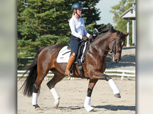
Main Photo: iStock/Asya Pozniak
Blogs & Podcasts

Advertisement

Related Articles


Advertisement

Featured Stories


Advertisement